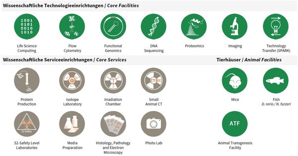
Core Facilites and Core Services
At the beginning of 2016, a “core” structure was put into effect that organized facility and service units as independent organizational entities from FLI’s research groups. A number of technology platforms (e.g. sequencing, mass spectrometry) grew out of individual methodological requirements for single research groups in the last years but developed into semiautonomous substructures. As consequence of re-focused research activities and the concomitant advent of new research groups at FLI, those units increasingly had to serve many FLI groups and collaborative research efforts in the Jena research area.
To accommodate this development and to increase efficiency as well as transparency for users, facility personnel and for administrative processes, it came natural to re-organize such activities into independent units as “FLI Core Facilities and Services” and to phase out infrastructures considered non-essential for FLI’s research focus (X-ray crystallography and NMR spectroscopy).
FLI’s Core Facilities (CF) are managed by a CF Manager and are each scientifically guided in their activities and development by an FLI Group Leader, as Scientific Supervisor. The animal facilities comprising fish, mouse and transgenesis are run separately, as they involve a more complex organizational structure. Basic Core Services (CS) are directly led by the Head of Core (HC), who in turn is supported by individual CS Managers.
All facilities and services, including animal facilities, have a valuable contribution to FLI’s research articles; e.g. from 2016–2018, to 54% of all peer reviewed research publications.
Overview Core Facilities and Core Services at FLI.
Publications
(since 2016)
2021
- Abundance and size of hyaluronan in naked mole-rat tissues and plasma.
Del Marmol D, Holtze S, Kichler N, Sahm A, Bihin B, Bourguignon V, Dogné S, Szafranski K, Hildebrandt TB, Flamion B
Sci Rep 2021, 11(1), 7951 - Mapping protein carboxymethylation sites provides insights into their role in proteostasis and cell proliferation.
Di Sanzo* S, Spengler* K, Leheis A, Kirkpatrick JM, Rändler TL, Baldensperger T, Dau T, Henning C, Parca L, Marx C, Wang ZQ, Glomb MA, Ori** A, Heller** R
Nat Commun 2021, 12(1), 6743 * equal contribution, ** co-senior authors - Molekularbiologische Beschreibung des Alterungsprozesses
Diekmann S, Grosse F, Hemmerich P, Pospiech H
In: Handbuch Alter und Altern, J.B. Metzler (edited by Fuchs M) 2021, 145–151, Springer, Heidelberg - Cell Type-Specific Role of RNA Nuclease SMG6 in Neurogenesis.
Guerra GM, May D, Kroll T, Koch P, Groth M, Wang** ZQ, Li TL, Grigaravičius** P
Cells 2021, 10(12), 3365 ** co-corresponding authors - Power and Weakness of Repetition - Evaluating the Phylogenetic Signal From Repeatomes in the Family Rosaceae With Two Case Studies From Genera Prone to Polyploidy and Hybridization ( Rosa and Fragaria).
Herklotz V, Kovařík A, Wissemann V, Lunerová J, Vozárová R, Buschmann S, Olbricht K, Groth M, Ritz CM
Front Plant Sci 2021, 12, 738119 - Alternative Animal Models of Aging Research
Holtze S, Gorshkova E, Braude S, Cellerino A, Dammann P, Hildebrandt TB, Hoeflich A, Hoffmann S, Koch P, Terzibasi Tozzini E, Skulachev M, Skulachev VP, Sahm A
Front Mol Biosci 2021, 8, doi: 10.3389/fmolb.2021.660959. - ATP/IL-33-triggered hyperactivation of mast cells results in an amplified production of pro-inflammatory cytokines and eicosanoids.
Jordan PM, Andreas N, Groth M, Wegner P, Weber F, Jäger U, Küchler C, Werz O, Serfling E, Kamradt T, Dudeck A, Drube S
Immunology 2021, 164(3), 541-54 - DNA-binding properties of the MADS-domain transcription factor SEPALLATA3 and mutant variants characterized by SELEX-seq.
Käppel S, Eggeling R, Rümpler F, Groth M, Melzer R, Theißen G
Plant Mol Biol 2021, 105(4-5), 543-57 - ATR regulates neuronal activity by modulating presynaptic firing.
Kirtay M, Sell J, Marx C, Haselmann H, Ceanga M, Zhou ZW, Rahmati V, Kirkpatrick J, Buder K, Grigaravicius P, Ori A, Geis** C, Wang** ZQ
Nat Commun 2021, 12(1), 4067 ** co-corresponding authors - Isolation, ‘omics characterization and organotypic culture of alveolar type II pulmonary epithelial cells
Li* H, Schütte* M, Bober* M, Kroll T, Frappart L, Bou About G, Lin YC, Sorg T, Herault Y, Wierling C, Rinner O, Lange Bodo MH, Ploubidou A
bioRxiv 2021, https://doi.org/10.1101/2021.08. * equal contribution


